On this page
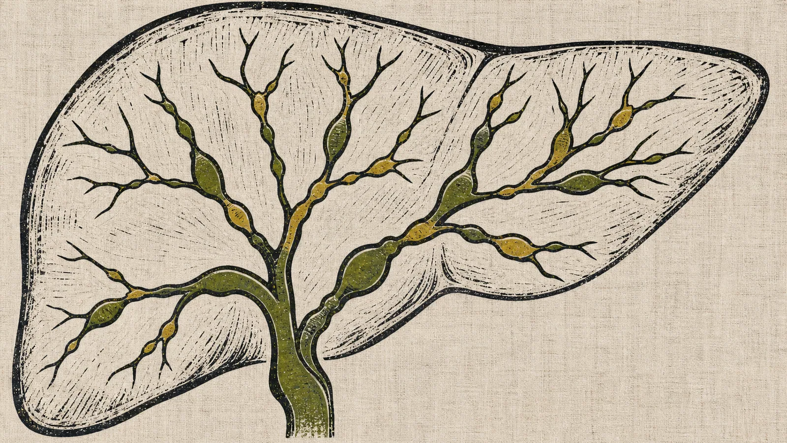
Primary sclerosing cholangitis (PSC) is a chronic liver disease in which the bile ducts, the channels that carry bile from the liver to the intestine, become inflamed and gradually fill with scar tissue (fibrosis). Over time that scarring forms narrowings, called strictures, that impede the flow of bile. Unlike most liver diseases, it is somewhat more common in men, and it has a very close relationship with inflammatory bowel disease, especially ulcerative colitis.
If you have been diagnosed with this condition, two things are worth knowing from the start. First, there is currently no medication proven to stop or reverse the course of PSC, so treatment focuses on managing complications and monitoring the associated risks. Second, the course varies a great deal from one person to another, and in many cases it progresses slowly over years.
What is its link with ulcerative colitis?
PSC and inflammatory bowel disease go hand in hand. Between half and four fifths of people with PSC also have inflammatory bowel disease, almost always ulcerative colitis and less often Crohn’s disease. The bowel involvement can cause very few symptoms, so a colonoscopy is recommended to examine the colon even when there are no digestive complaints.
The relationship also works the other way around, though with much lower figures: only a small fraction of people with ulcerative colitis develop PSC. The two diseases can appear at different times, and the activity of one does not predict the activity of the other.
Does it cause symptoms?
In its early stages PSC is usually asymptomatic. It is often suspected because of an incidental finding: abnormal liver tests in a person with ulcerative colitis, in particular elevation of alkaline phosphatase and GGT, which reflect a cholestatic pattern (difficulty in bile flow).
When symptoms do appear, the most common are itching (pruritus) and fatigue. Some patients have episodes of fever, jaundice and right-sided abdominal pain, which correspond to cholangitis, that is, infection of the bile ducts. In advanced stages, the complications of cirrhosis may appear, such as ascites, varices or encephalopathy.
How is it diagnosed?
The diagnosis rests on three pillars: the cholestatic pattern in the blood, imaging of the bile ducts, and exclusion of other causes.
- Blood tests. They show the cholestatic pattern described above. About half of patients have antineutrophil cytoplasmic antibodies with a perinuclear pattern (p-ANCA), but no blood test alone confirms the diagnosis.
- MR cholangiography (MRCP). This is now the test of choice. It is a magnetic resonance scan that shows the biliary tree without introducing any instruments, and it reveals the characteristic strictures and dilatations that alternate along the ducts. It has almost entirely replaced endoscopic retrograde cholangiography (ERCP) for diagnosis.
- ERCP. Endoscopic retrograde cholangiopancreatography is now reserved for uncertain cases or, above all, when a stricture needs to be treated or samples taken, because it is a procedure that carries risks.
A liver biopsy is not necessary for the diagnosis in most cases. It is reserved for special situations, such as suspicion of small-duct PSC (which is not visible on imaging) or of overlap with autoimmune hepatitis. When taken, the typical finding is fibrosis arranged in concentric layers around the small bile ducts, the so-called “onion-skin” lesion.
The differential diagnosis must include causes of secondary sclerosing cholangitis, such as cholangiopathy related to infection or ischemia, and IgG4-associated cholangitis (formerly grouped with autoimmune pancreatitis), which is important to distinguish because it does respond to treatment with corticosteroids.
Is there any treatment?
This is the hardest question to answer, because today no medication has been shown to modify the course of the disease or to improve survival. Management is organized on two fronts: treating complications and monitoring the associated cancers.
Regarding ursodeoxycholic acid (UDCA), a bile acid widely used in other cholestatic diseases, the evidence is clear on one point. A controlled trial using high doses (28 to 30 mg/kg per day) was stopped early because the treated group had more serious complications than the placebo group. For that reason, current AASLD and EASL guidelines do not recommend high-dose UDCA. Its use at low or moderate doses improves blood tests, but does not change the prognosis, and the guidelines do not routinely endorse it. Any prescription should be made by your specialist.
Management of complications includes:
- Dominant stricture. When a significant stricture blocks the flow of bile and causes jaundice or cholangitis, it can be treated endoscopically by ERCP. Balloon dilatation is preferred and stents are avoided when possible. Any dominant stricture requires ruling out a bile duct cancer.
- Cholangitis. Episodes of biliary infection are treated with antibiotics. Recurrent bacterial cholangitis is one of the reasons to consider transplantation.
- Itching. There are specific treatments for the itching, which can be very distressing. See the management of itching.
- Nutritional and bone problems. Cholestasis impairs absorption of the fat-soluble vitamins (A, D, E and K) and favors osteoporosis, so these aspects should be monitored.
Cancer risk and surveillance
PSC raises the risk of several cancers, which is why surveillance is a central part of care.
- Cholangiocarcinoma (bile duct cancer). This is the most feared complication. The lifetime risk is considerable and diagnosis is difficult, because it is easily confused with the strictures that belong to the disease itself. Guidelines recommend surveillance with imaging (MR cholangiography or ultrasound) and the CA 19-9 marker, generally once a year. It should be suspected with unexplained weight loss, a rapid deterioration or a new dominant stricture.
- Gallbladder cancer. Gallbladder polyps in PSC carry a higher risk of being malignant, so the gallbladder is monitored and sometimes removed.
- Colorectal cancer. In those who also have ulcerative colitis, the risk of colon cancer is higher than with colitis alone, so surveillance colonoscopy with biopsies is recommended, usually every one to two years.
Transplant and prognosis
Liver transplant is the treatment of choice when the disease progresses toward cirrhosis with complications. Outcomes of transplantation in PSC are good, although the disease can recur in the transplanted liver in roughly one in five patients. Recurrent bacterial cholangitis and, in highly selected cases and under strict protocols, early-stage cholangiocarcinoma, can be indications for transplantation.
The prognosis varies widely. PSC is generally a slowly progressive disease that unfolds over years, and survival depends on the pace of fibrosis, the appearance of complications and the risk of cancer. This is why follow-up with a specialized liver team, together with the team managing the bowel disease, is what helps most.
See also
References
- Bowlus CL, et al. AASLD practice guidance on primary sclerosing cholangitis and cholangiocarcinoma. Hepatology. 2023;77(2):659-702.
- European Association for the Study of the Liver. EASL Clinical Practice Guidelines on sclerosing cholangitis. J Hepatol. 2022;77(3):761-806.
- von Seth E, et al. Primary sclerosing cholangitis. Lancet. 2026;407(10538):1549-1569.
- Lindor KD, et al. High-dose ursodeoxycholic acid for the treatment of primary sclerosing cholangitis. Hepatology. 2009;50(3):808-814.
Related articles
Autoimmune hepatitis
Autoimmune hepatitis happens when the immune system attacks the liver itself. What it is, how it is diagnosed and how it is treated with corticosteroids and azathioprine.
Primary biliary cholangitis (formerly primary biliary cirrhosis)
Primary biliary cholangitis (formerly primary biliary cirrhosis) is an autoimmune disease of the bile ducts. What it is, how it is diagnosed and how it is treated.
Hepatocellular adenoma: what it means and when it needs treatment
A hepatocellular adenoma is benign, but some may bleed or grow. Learn how hormones, obesity, pregnancy and tumor size affect treatment and follow-up.