Cholangiocarcinoma is a cancer that begins in the bile ducts, the channels that carry bile from the liver to the intestine. It is less common than hepatocellular carcinoma, but it tends to be more aggressive and is often discovered because it causes obstructive jaundice, a yellowing of the skin and eyes from blocked bile flow.
If you or a family member has just been given this diagnosis, three things are worth knowing at the outset. First, not all cholangiocarcinomas are alike, their location changes the prognosis and the treatment. Second, when the tumor can be removed with surgery, cure is possible. And third, in recent years the treatment of advanced cases has changed, with the addition of immunotherapy and of targeted therapies aimed at specific tumor mutations.
What is cholangiocarcinoma?
Bile is produced in the liver and travels through a network of ducts that merge into the main bile duct, which empties into the intestine. Cholangiocarcinoma is the cancer that arises from the lining of those ducts. It is distinct from gallbladder cancer and from ampullary tumors (cancer of the ampulla of Vater), which are neighboring but different diseases.
Depending on where it starts, it is classified into three types, and this distinction matters because it changes the symptoms, the surgery and the prognosis:
- Intrahepatic (about 10 to 15% of cases): arises in the small ducts inside the liver. It is usually found as a mass on imaging rather than through jaundice. Its incidence is rising worldwide.
- Perihilar or hilar (around 50 to 60%): begins where the ducts leave the liver and join together. When it involves that junction it is called a Klatskin tumor. This is the type that most often causes early jaundice.
- Distal (about 20 to 25%): located in the final part of the bile duct, close to the pancreas and the intestine.
Risk factors
In most people cholangiocarcinoma appears without an identifiable cause. The typical age at presentation is between 50 and 70, though it can occur earlier in those with risk factors. The main ones are:
- Primary sclerosing cholangitis: the best-recognized risk factor. This disease, which inflames and narrows the bile ducts and is often associated with ulcerative colitis, markedly increases lifetime risk.
- Bile duct cysts and malformations: choledochal cysts, Caroli disease and congenital hepatic fibrosis raise the risk.
- Stones in the intrahepatic bile ducts (hepatolithiasis): common in Asia, where it is also known as oriental cholangiohepatitis.
- Bile duct parasites: chronic infection with the liver flukes Clonorchis sinensis and Opisthorchis viverrini, endemic in Southeast Asia, is a well-established cause.
- Chronic liver disease and cirrhosis: hepatitis B, hepatitis C and cirrhosis of any cause raise the risk of the intrahepatic form in particular. Diabetes, obesity and alcohol use have also been linked.
Symptoms
Symptoms depend on where the tumor is. When it blocks the bile duct, bile cannot drain and the following appear:
- Jaundice: yellowing of the skin and the whites of the eyes.
- Dark urine: urine the color of tea or cola.
- Pale stools and itching (pruritus) from bile salts building up in the skin.
Other common symptoms are unexplained weight loss, fatigue and a dull ache or discomfort in the right side of the abdomen. Intrahepatic tumors can grow considerably before causing symptoms, because they do not always block the main duct.
Diagnosis
The workup combines imaging, blood tests and, when possible, obtaining samples of the tumor.
- Imaging: ultrasound is usually the first test, but the evaluation is completed with CT and, above all, with MR cholangiography (an MRI focused on the bile ducts), which is today the best method to define the tumor’s anatomy and plan surgery. A PET scan helps look for metastases.
- Tumor markers: CA 19-9 may be elevated and is useful for suspicion and follow-up, although it also rises in benign obstructions and other cancers, so it is not enough on its own. It is sometimes combined with carcinoembryonic antigen (CEA).
- Cholangiography and biopsy: through endoscopic retrograde cholangiopancreatography (ERCP), often combined with endoscopic ultrasound, biopsies or brushings of the bile duct can be taken and, if needed, a stent can be placed to relieve the obstruction.
Telling a cholangiocarcinoma apart from a benign narrowing is still difficult in many cases, especially in people with primary sclerosing cholangitis, where diagnosis requires close follow-up by an experienced team.
Treatment
Treatment depends on whether the tumor can be removed completely.
Surgery. This is the only treatment that can cure, and it is always pursued when the tumor is resectable. The operation varies by location and should ideally be performed at a center experienced in hepatobiliary surgery, within a multidisciplinary team. Unfortunately, many patients reach diagnosis when the tumor is already advanced and inoperable.
Liver transplant. In highly selected cases of perihilar cholangiocarcinoma, especially those linked to primary sclerosing cholangitis, liver transplant preceded by chemotherapy and radiation (a protocol developed by the Mayo Clinic) can offer good results. It is an option for few patients and only at centers with specific programs.
Advanced disease. When the tumor cannot be operated on or has already spread, the goal is to prolong life with good quality. There have been meaningful advances here:
- Chemotherapy with immunotherapy: for more than a decade the standard was chemotherapy with gemcitabine and cisplatin. The TOPAZ-1 trial showed that adding the immunotherapy drug durvalumab to that chemotherapy improves survival, and this combination is now the new first-line treatment in many countries.
- Targeted therapies based on mutations: in intrahepatic cholangiocarcinoma it is increasingly important to study the tumor’s molecular profile. When an FGFR2 gene fusion is present, drugs such as pemigatinib are effective. When there is an IDH1 mutation, ivosidenib offers benefit. These therapies are used mainly when the initial chemotherapy stops working.
- Managing the bile duct obstruction: placing a stent by endoscopy or, less often, by surgery, relieves jaundice and itching and improves quality of life.
The availability of immunotherapy and targeted therapies is still uneven and depends on each person’s health system. The decision to test the tumor’s mutations and to prescribe these drugs should always be made by your treating team.
See also
References
- Valle JW, Kelley RK, Nervi B, Oh DY, Zhu AX. Biliary tract cancer. Lancet. 2021;397(10272):428-444.
- Oh DY, et al. Durvalumab plus gemcitabine and cisplatin in advanced biliary tract cancer (TOPAZ-1). NEJM Evid. 2022;1(8):EVIDoa2200015.
- Abou-Alfa GK, et al. Pemigatinib for previously treated, locally advanced or metastatic cholangiocarcinoma (FIGHT-202): a multicentre, open-label, phase 2 study. Lancet Oncol. 2020;21(5):671-684.
- Abou-Alfa GK, et al. Ivosidenib in IDH1-mutant, chemotherapy-refractory cholangiocarcinoma (ClarIDHy): a phase 3 study. Lancet Oncol. 2020;21(6):796-807.
Related articles
Hepatocellular adenoma: what it means and when it needs treatment
A hepatocellular adenoma is benign, but some may bleed or grow. Learn how hormones, obesity, pregnancy and tumor size affect treatment and follow-up.
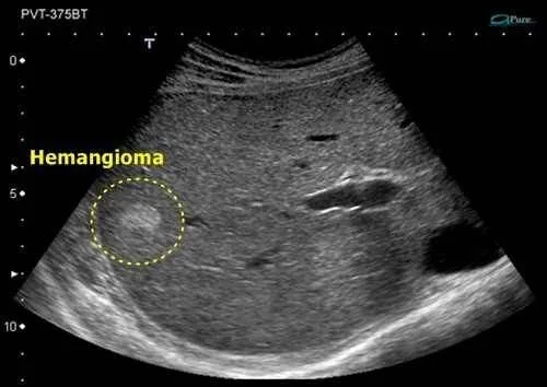
Liver hemangioma
A liver hemangioma is the most common benign liver tumor. It is almost always an incidental finding, causes no symptoms and needs no treatment.
Focal nodular hyperplasia of the liver (FNH)
Focal nodular hyperplasia is a benign liver tumor, most common in young women. It is almost always an incidental finding, does not become cancer and needs no treatment.