On this page
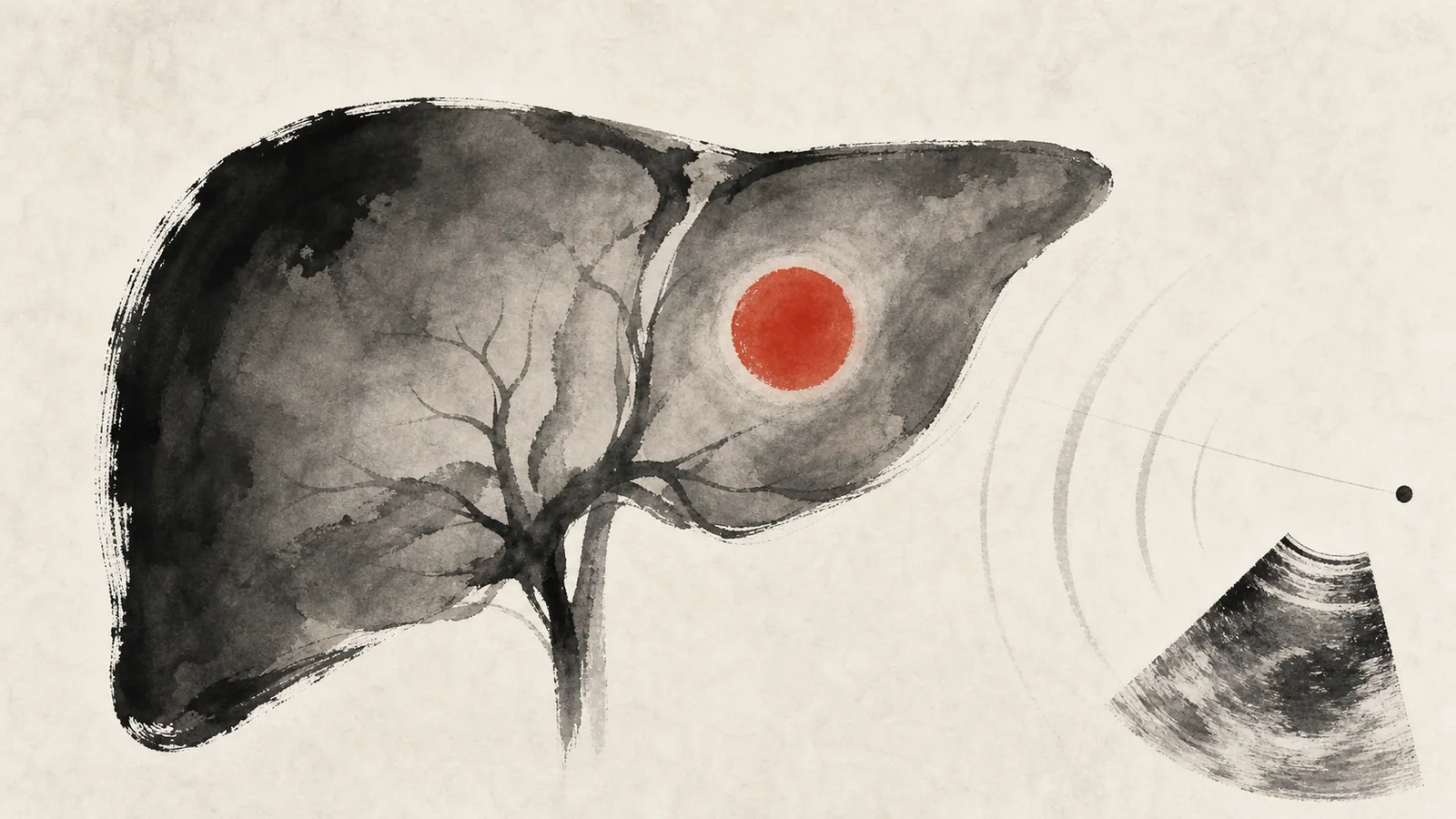
Hepatocellular carcinoma (HCC), also called hepatoma or liver cancer, is the most common primary cancer of the liver, meaning the one that begins in the liver tissue itself. It almost always arises in a liver that is already damaged, particularly in a cirrhotic liver, so in most cases it is possible to know in advance who should be monitored.
The good news is that a large share of hepatocellular carcinoma can be prevented by treating the underlying cause of liver damage, and that when it is detected early, while the liver is still working well, there are treatments that can cure it. This is why people with cirrhosis should have regular checkups, even when they feel healthy.
What is it and why does it appear?
Hepatocellular carcinoma originates in hepatocytes, the main cells of the liver. It rarely arises in a healthy liver. In more than 80% of cases it develops on top of cirrhosis, regardless of the cause. The highest-risk situations are:
- Cirrhosis from chronic hepatitis B or hepatitis C.
- Cirrhosis from alcohol.
- Cirrhosis from fatty liver associated with metabolic dysfunction (MASLD), an increasingly common cause.
- Hemochromatosis and other chronic liver diseases.
Hepatitis B deserves a separate mention, because it can cause hepatocellular carcinoma even before the patient develops cirrhosis.
Can it be prevented?
To a large extent, yes. The most effective prevention is to avoid or treat the liver disease that leads to the cancer:
- Hepatitis B vaccine. It is the only vaccine that prevents a human cancer. It has clearly reduced the risk of hepatocellular carcinoma wherever it has been implemented.
- Treating hepatitis C. Direct-acting antivirals cure the infection in the great majority of patients and lower the risk of cancer, although in those who already have cirrhosis the risk is not fully eliminated.
- Controlling hepatitis B with antivirals when indicated.
- Avoiding alcohol and managing fatty liver, diabetes and obesity.
Early detection: ultrasound every 6 months
The challenge is not so much diagnosing hepatocellular carcinoma once it causes symptoms, but finding it at an early stage, when it can still be cured. This is the purpose of surveillance (screening), aimed at the people at greatest risk.
Surveillance is recommended for all patients with cirrhosis, whatever the cause, and for some patients with chronic hepatitis B without cirrhosis. The accepted strategy is:
- Abdominal ultrasound every 6 months. This is the core test: non-invasive, no radiation and low cost.
- Alpha-fetoprotein (AFP) in the blood, usually added to the ultrasound. It is a marker with limitations, since it can be normal in small tumors and rise for other reasons, so it never replaces imaging.
When the ultrasound shows a suspicious nodule, the next step is a contrast-enhanced scan to confirm the diagnosis.
Diagnosis by imaging
Hepatocellular carcinoma has a feature that makes it easier to diagnose: it is a highly vascular tumor. On contrast studies it takes up contrast quickly in the arterial phase and then “washes out” in the later phases, a fairly characteristic pattern.
- Contrast-enhanced computed tomography (CT), in several phases.
- Contrast-enhanced magnetic resonance imaging (MRI), with sensitivity equal to or greater than CT, especially useful in the cirrhotic liver.
In patients with cirrhosis, radiologists classify nodules using the LI-RADS system, which grades the degree of suspicion for cancer. Thanks to these criteria, in many cases the diagnosis is made by imaging alone, without a biopsy. A liver biopsy is reserved for when doubt persists or the nodule appears in a non-cirrhotic liver, since it carries risks such as bleeding and, very rarely, tumor seeding along the needle track.
Does it cause symptoms?
In early stages, usually not. That is why surveillance matters so much. When the tumor is advanced, nonspecific symptoms may appear, such as weight loss, pain in the right side of the abdomen, early fullness or a palpable mass. Sometimes it presents as a sudden decompensation in a patient with previously stable cirrhosis, with jaundice, ascites or bleeding.
Treatment by stage
Because hepatocellular carcinoma usually sits on top of a cirrhotic liver, the treatment decision depends on three things at once: the features of the tumor (number, size, extent), how well the liver is working, and the patient’s general condition. These variables are combined in the BCLC classification (Barcelona Clinic Liver Cancer), the most widely used framework worldwide to guide stage-based therapy.
The decision should always be made by an experienced multidisciplinary team that brings together hepatologists, surgeons, radiologists and oncologists. The main options are:
Surgical resection
Removing the part of the liver that contains the tumor. It is a curative-intent option in patients with a single tumor, good liver function and no significant portal hypertension. Only a minority of patients meet these conditions.
Liver transplant
A liver transplant is the ideal treatment when feasible, because it removes the tumor and cures the cirrhosis at the same time. It is offered to patients whose tumor meets the Milan criteria:
- A single tumor up to 5 cm, or
- Up to 3 tumors, none larger than 3 cm.
In these cases survival after transplant is excellent. Because the wait for an organ can be long, “bridging” therapies such as ablation or chemoembolization are often used to control the tumor until transplant.
Local ablation
Destroying the tumor with heat delivered through a needle, either radiofrequency ablation or microwave ablation. It is a curative option for small tumors, especially when surgery is not possible, and also serves as a bridge to transplant. The older ethanol injection has become secondary because it is less effective.
Chemoembolization (TACE)
In transarterial chemoembolization, chemotherapy is injected together with an agent that blocks the artery feeding the tumor, through a catheter. It is the treatment of choice for intermediate tumors, larger or multiple but still confined to the liver. It can cause a post-embolization syndrome, with transient fever and abdominal pain.
Systemic therapy
When the tumor is advanced, has spread outside the liver or invades the vessels, treatment becomes systemic, that is, with medications that act throughout the body. This is where practice has changed the most in recent years.
For a long time the only option was sorafenib. Today, in patients with good liver function, the first line is combination immunotherapy:
- Atezolizumab plus bevacizumab, which in a phase 3 trial showed longer survival than sorafenib.
- Durvalumab plus tremelimumab (the STRIDE regimen), another combination that also outperformed sorafenib in survival.
Sorafenib and other oral drugs (such as lenvatinib) remain useful in patients who cannot receive immunotherapy or as later lines. The choice of regimen depends on liver function and each patient’s other conditions, so it must be individualized.
What is the prognosis?
It depends above all on the stage at which it is detected. A hepatocellular carcinoma found early, in a liver that still works well, can be treated with curative intent and has a good prognosis. When it is discovered at an advanced stage, the options are more limited. This is why hepatitis B vaccination, treatment of viral hepatitis and ultrasound surveillance in people with cirrhosis are so important: they are the best way to change the prognosis.
See also
References
- Singal AG, et al. AASLD Practice Guidance on prevention, diagnosis, and treatment of hepatocellular carcinoma. Hepatology. 2023;78(6):1922-1965.
- Reig M, et al. BCLC strategy for prognosis prediction and treatment recommendation: The 2022 update. J Hepatol. 2022;76(3):681-693.
- Finn RS, et al. Atezolizumab plus Bevacizumab in Unresectable Hepatocellular Carcinoma. N Engl J Med. 2020;382(20):1894-1905.
- Abou-Alfa GK, et al. Tremelimumab plus Durvalumab in Unresectable Hepatocellular Carcinoma. NEJM Evid. 2022;1(8):EVIDoa2100070.
- Llovet JM, et al. Hepatocellular carcinoma. Nat Rev Dis Primers. 2021;7(1):6.
Related articles
Compensated cirrhosis: follow-up and care
Compensated cirrhosis can remain stable for years. Learn which regular follow-up checks help prevent decompensation and detect liver cancer early.

How to choose a good hepatologist in Chile
Verifiable criteria for choosing a hepatologist in Chile: professional registration, training, relevant experience, clinical network, communication and follow-up.
Causes of liver cirrhosis
The causes of cirrhosis, from the most to the least common, and why identifying and treating the cause early can stop and even reverse liver damage.