On this page
If an ultrasound or a CT scan has found a liver hemangioma, the underlying news is reassuring: it is the most common benign tumor of the liver, it is not a cancer, and it does not turn into cancer. In the vast majority of people it causes no symptoms, needs no treatment, and does not even require lifelong follow-up.
A hemangioma, also called a cavernous hemangioma, is not really a tumor in the usual sense but a malformation of blood vessels within the liver, a kind of tangle of small vessels present from birth. Its only practical importance is usually to tell it apart, with confidence, from other lesions, something that imaging does well today. Only large hemangiomas or those that cause symptoms need a more detailed evaluation.
How common is it?
Very common. An estimated 0.4 to 20% of people have a liver hemangioma, depending on how it is looked for, and it is more common in women. It is usually found by chance on imaging ordered for another reason, most often between the ages of 30 and 50, although it can appear at any age. Most are small and solitary, but having more than one is not unusual.
Why does it happen?
The cause is not well understood. It is considered a congenital vascular malformation, meaning it is present from before birth, that grows through slow dilation of its vessels rather than through the multiplication of cells as a cancer does. This is why its behavior is so stable.
Female hormones (estrogens) have been proposed to influence its growth, based on reports of enlargement during pregnancy or with contraceptives. The evidence is limited, and not all hemangiomas respond to hormones, so in practice this rarely changes the recommendations.
Does it cause symptoms?
Almost never. The great majority of hemangiomas produce no discomfort and are an incidental finding. It is very unlikely that a lesion smaller than 4 to 5 cm will cause symptoms.
When symptoms do occur, the most common is pain or discomfort in the upper abdomen, sometimes with early fullness or a sense of a lump. It is wise to be cautious about blaming the pain on the hemangioma, because in more than half of cases the pain has another cause, often irritable bowel syndrome. Rarely, a large hemangioma can develop a thrombosis or bleeding within the lesion, which appears as sudden abdominal pain.
Kasabach-Merritt syndrome is a rare and distinct situation, seen with giant hemangiomas in infants, that causes clotting abnormalities. It is not what happens with the usual adult hemangioma.
Can it grow or rupture?
Generally, no. Follow-up studies show that more than 80% of hemangiomas keep the same size over the years. Only a small proportion grow slowly, and spontaneous rupture is exceptional, essentially limited to large lesions (over 5 cm) in a peripheral location. This very low risk is the reason a hemangioma is almost never operated on for its size alone.
How is it diagnosed?
A hemangioma has very suggestive features on imaging, which often allows it to be confirmed without a biopsy.
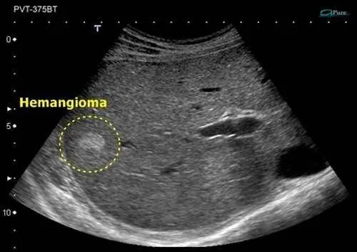
- Ultrasound: it usually appears as a well-defined, hyperechoic (“brighter”) lesion compared with the liver. When there is fatty liver, it can look the opposite, darker than the surrounding tissue. In a person without cirrhosis, without a known cancer, and with a small, typical lesion, no further tests are sometimes needed.
- Magnetic resonance imaging (MRI): this is the reference test. The hemangioma looks hypointense on T1 sequences and strongly hyperintense on T2, and with contrast it shows a very characteristic filling pattern that begins at the edges and moves toward the center. This behavior allows a highly confident diagnosis.
- Contrast-enhanced CT: it shows the same pattern of progressive filling from the periphery toward the center and is a good alternative when MRI is not available.
A biopsy is almost never recommended when a hemangioma is suspected, because imaging is usually enough and puncturing a vessel-filled lesion carries a risk of bleeding.
How is it told apart from other liver lesions?
The main goal of the workup is to confirm that the lesion is benign and to distinguish it from others. The ones most often considered in the differential are focal nodular hyperplasia and hepatic adenoma, both also benign, along with a simple liver cyst. In people with cirrhosis or with a known cancer elsewhere, the workup is more careful in order to rule out a hepatocellular carcinoma or a metastasis. In most cases, the MRI pattern settles the question.
Does it need treatment?
For the vast majority of people, no. A small, typical, symptom-free hemangioma needs neither treatment nor imaging follow-up, and it places no restrictions on daily life. You can exercise, travel, and live normally.
Large hemangiomas are sometimes checked with a follow-up image at a few months or at one year to confirm they remain stable. If they are clearly stable, there is no need to keep monitoring them for life.
Surgery is reserved for the few cases with persistent symptoms clearly attributable to the lesion, or with complications. Before operating, it is essential to confirm that the pain truly comes from the hemangioma, because when it does not, the discomfort continues after surgery. The operation may be a resection or an enucleation (removing the lesion while sparing the rest of the liver), and in very selected giant hemangiomas there is the option of a liver transplant. Other techniques, such as embolization, are used in specific situations.
Regarding pregnancy and estrogen-containing contraceptives, there is no reason to forbid them because of a small hemangioma. If the lesion is large, it is worth discussing with your physician, although most women go through pregnancy without problems.
See also
References
- Frenette C, et al. ACG Clinical Guideline: Focal Liver Lesions. Am J Gastroenterol. 2024;119(7):1235-1271.
- European Association for the Study of the Liver. EASL Clinical Practice Guidelines on the management of benign liver tumours. J Hepatol. 2016;65(2):386-398.
- Aziz H, et al. A Comprehensive Review of Hepatic Hemangioma Management. J Gastrointest Surg. 2022;26(9):1998-2007.
- Reguram R, et al. Practical approach to diagnose and manage benign liver masses. Hepatol Commun. 2024;8(11):e0560.
Related articles

Hepatocellular adenoma: what it means and when it needs treatment
A hepatocellular adenoma is benign, but some may bleed or grow. Learn how hormones, obesity, pregnancy and tumor size affect treatment and follow-up.
Focal nodular hyperplasia of the liver (FNH)
Focal nodular hyperplasia is a benign liver tumor, most common in young women. It is almost always an incidental finding, does not become cancer and needs no treatment.

Polycystic liver disease
Polycystic liver disease means multiple cysts in the liver of genetic origin. It is usually symptom-free and liver function is preserved. What it is and when it is treated.