On this page
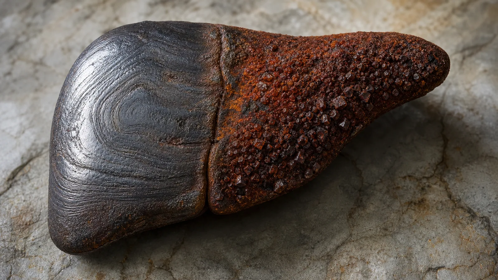
Hemochromatosis is a condition in which the body absorbs and stores too much iron. Over the years, that excess is deposited in various organs, above all the liver, and can damage them. It is one of the most common genetic diseases in people of European ancestry, and the good news is that, when caught early, it is treated simply and with an excellent prognosis.
If you were told your ferritin is high, it is natural to feel alarmed when reading about hemochromatosis. It helps to keep calm: most elevated ferritin results do not reflect true iron overload but other, far more common causes, such as fatty liver disease. Below we explain how they are told apart.
Why does iron build up?
The body has no efficient way to get rid of excess iron, so it carefully regulates how much it absorbs from the gut. In hemochromatosis that regulation fails and iron keeps being absorbed even when stores are already full. The excess is deposited in the liver, heart, pancreas and joints, and over time causes damage, largely through oxidative stress (free radicals).
The most common form is hereditary hemochromatosis caused by a mutation in the HFE gene. It is inherited in an autosomal recessive way, meaning you need to inherit the mutation from both parents to be at risk of developing the disease. The mutation responsible in the vast majority of cases is called C282Y (when inherited in both copies of the gene, known as homozygosity). A second variant, H63D, causes iron overload only in some people, especially when combined with C282Y (compound heterozygote).
It is important to know that not everyone with the mutation develops the disease. Penetrance is incomplete: many C282Y homozygotes never accumulate enough iron to become ill. In addition, HFE mutations explain most cases in populations of European ancestry, but far fewer in Asian and Latin American populations. In Chile a substantial share of patients with iron overload do not carry the typical HFE mutations, so a negative genetic test does not rule out the problem.
There are other causes of iron accumulation that are not hereditary hemochromatosis, such as repeated red blood cell transfusions or certain anemias. In those cases the term used is usually hemosiderosis or secondary iron overload.
What symptoms does it cause?
At the beginning, none. Hemochromatosis is silent for years, which is why most diagnoses today are made before symptoms appear, prompted by abnormal blood tests or family screening. The classic “bronze diabetes” with cirrhosis described in old textbooks is now rare, because the disease is detected far earlier.
When iron does damage the organs, it can show up as:
- Fatigue and low energy, often the first symptom.
- Joint pain and stiffness (arthropathy), typically in the knuckles of the index and middle fingers.
- Abnormal liver tests, an enlarged liver and, in advanced cases, cirrhosis and even liver cancer.
- Diabetes, from iron deposits in the pancreas.
- Heart problems, from rhythm disturbances to heart failure.
- Reduced sex drive and impotence, from lower sex hormones.
- Darkening of the skin (a grayish or bronze color).
Drinking alcohol and infection with the hepatitis C or hepatitis B viruses speed up liver damage. Once cirrhosis is present, the risk of liver cancer is higher than in other causes of cirrhosis, so these patients need regular monitoring.
How is it diagnosed?
The workup starts with simple blood tests, the so-called iron studies:
- Transferrin saturation. This is the earliest marker and the most useful for suspecting true iron overload. A saturation above 45% warrants investigation; in hemochromatosis it is usually markedly elevated.
- Ferritin. This reflects the body’s iron stores, but it also rises with inflammation, fatty liver, alcohol use and other causes. That is why a high ferritin alone is not enough to diagnose hemochromatosis.
When both tests are persistently elevated, the next step is the HFE genetic test, which looks for the C282Y and H63D mutations. If it confirms C282Y homozygosity, the diagnosis is established. If the genetic study does not explain the overload, or to measure how much iron has accumulated, liver MRI is used, a noninvasive test that estimates iron content without needing to sample the liver.
A liver biopsy is no longer needed for diagnosis in most cases. It is reserved for specific situations, mainly to assess the degree of fibrosis or when ferritin is very high (for example, above 1,000 ng/mL), which is when the risk of cirrhosis increases.
High ferritin is not the same as hemochromatosis
This is a very common source of confusion and worth pausing on. The most common cause of elevated ferritin today is not hemochromatosis but fatty liver disease, which affects one in four people. In fatty liver, ferritin rises as a reflection of inflammation and the metabolic syndrome, but there is no true iron overload in the body.
The key to telling them apart is the transferrin saturation: in hemochromatosis it is high, while in fatty liver it is usually normal. So when ferritin is elevated, the first step is to look at the transferrin saturation and to consider common causes such as fatty liver or alcohol use before thinking of hemochromatosis.
Treatment
The treatment is as simple as it is effective: remove the excess iron by drawing blood. This is phlebotomy, very much like donating blood. About 450 to 500 mL are removed, and as the body replaces that blood it mobilizes the iron stored in the organs to make new red blood cells.
- Depletion phase. At first phlebotomies are frequent, usually once a week, until iron stores fall. This can take several months.
- Maintenance phase. Afterward only a few phlebotomies a year are needed to keep ferritin in the low range.
In the few people who cannot tolerate phlebotomy because of another illness, iron chelating drugs can be used; they bind iron and help remove it, although they are less practical and are second line.
Beyond treatment, it is advisable to avoid alcohol, not take iron or high-dose vitamin C supplements, and keep healthy eating habits to avoid adding damage from fatty liver.
What is the prognosis?
Very good when the diagnosis is timely. If iron is removed before cirrhosis develops, life expectancy is essentially normal and many symptoms improve. By contrast, once cirrhosis is established, treatment controls the disease but the liver damage does not reverse, and the risk of complications and liver cancer remains. That is why early diagnosis makes all the difference.
Family screening is essential
Because hereditary hemochromatosis is genetic, when one person is diagnosed it is advisable to screen their first-degree relatives (siblings, parents and adult children) with iron studies and, when appropriate, the genetic test. Detecting the overload in a relative who still has no symptoms allows timely treatment and can prevent organ damage entirely.
See also
References
- Bacon BR, et al. Diagnosis and management of hemochromatosis: 2011 practice guideline by the American Association for the Study of Liver Diseases. Hepatology. 2011;54(1):328-343.
- European Association for the Study of the Liver. EASL Clinical Practice Guidelines on haemochromatosis. J Hepatol. 2022;77(2):479-502.
- European Association for the Study of the Liver. EASL clinical practice guidelines for HFE hemochromatosis. J Hepatol. 2010;53(1):3-22.
- Adams PC, Jeffrey G, Ryan J. Haemochromatosis. Lancet. 2023;401(10390):1811-1821.
- Kowdley KV, et al. ACG Clinical Guideline: Hereditary Hemochromatosis. Am J Gastroenterol. 2019;114(8):1202-1218.
Related articles

Gilbert's syndrome
Gilbert's syndrome is a very common, harmless inherited condition that mildly raises bilirubin. It is not a liver disease and needs no treatment.
Hepatocellular adenoma: what it means and when it needs treatment
A hepatocellular adenoma is benign, but some may bleed or grow. Learn how hormones, obesity, pregnancy and tumor size affect treatment and follow-up.

The first months after a liver transplant: care and warning signs
A practical guide to the first months after a liver transplant: follow-up, immunosuppressants, safe recovery, infection prevention and warning signs.